
Out-patient Cone Beam Computerized Tomography
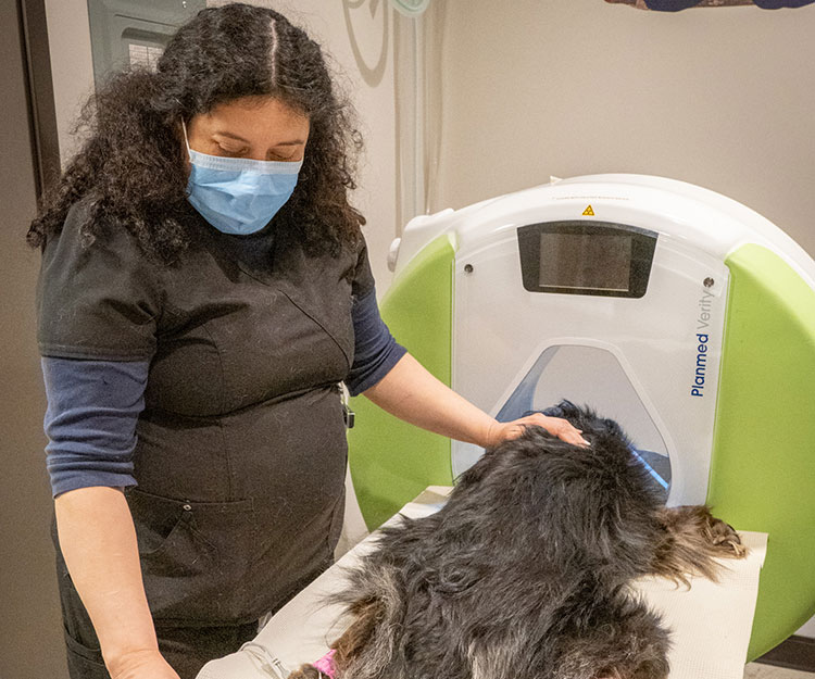
At Oregon Veterinary Dental Specialists we diagnose and treat severe oro-facial disease and trauma. A big part of our maxillofacial diagnostics is the use of Cone Beam Computerized Tomography (CBCT) technology. It provides instant 3-D viewing and high definition imaging of the head. It can also be useful for imagining the neck and extremities (areas we do not treat).
OVDS would like to offer this technology to our referring veterinary community to aid in the diagnosis of difficult conditions. For example:
- Nasal cavity (chronic sinusitis, discharge, airflow obstruction, etc.)
- Ears – tympanic bulla, inner, middle and external ear anatomy (chronic infection, polyps, neoplasia, etc.)
- Neck – bony trauma, subluxation, stenotic canal, etc. (Note MRI is better for soft tissue, CT is good for bone).
- Extremity lumps, bumps & lameness (elbow/stifle and distal anatomy).
What you can expect:
An outpatient CBCT is for patients being referred for CT, with an interpretation by a board certified radiologist only. All outpatient CTs will have a pre anesthetic exam with one of our surgery doctors the morning of the CT. This doctor will work with you to make sure you get the best diagnostic images possible. All CTs are sent to a board certified radiologist for interpretation and result times may vary. Our doctor can be available for tissue collection if requested ahead of time.
Pre-visit request:
- Current lab work within 3 months
- The last year of medical records
- Outpatient CT form completed by rDVM
- All information is required 2 days prior to the procedure.
***Urgent cases can be accommodated as needed. Please call the office for more information.